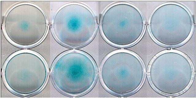
Micromass is based on the technique devised by Umansky (1966) to study the development and differentiaiton of cultured chick embryo limb cells. It was shown that the undifferentiated mesenchyme cells of limb buds will form foci of differentiating chondrocytes in micromass culture.
The present method is based on detecting the ability of a particular chemical to inhibit the formation of foci. Thus, positive chemicals will reduce the number of foci, or the number of cells within the foci.
The experimental system is the undifferentiated limb bud cells of a rat embryo. Embryos are obtained from Wistar rats on day 14 of gestation and the limb buds are isolated. Single cell suspension is prepared by trypsin action. The subsequent step, spotting of cells into 96 well plates is the most critical: place with care the spot in the center of the well and make the volume and number of cells within the spot as consistent as possible. Place the 96 well plates into incubator then add medium with or without test chemical and leave them for 5 days. At the and, the total number of viable cells (i.e. IC50: 50% inhibition of cell viability and growth) and of differentiated cells (i.e. ID50: 50% inhibition of cell differentiation) and number of foci were determined. The cells can be made visible by alcian blue staining (cartilage-specific proteoglycan stain). The endpoint is IC50 and ID50.
